CALEC Surgery: New Hope for Eye Damage Repair
In the realm of ocular health, CALEC surgery marks a significant breakthrough in the treatment of corneal damage, offering a glimmer of hope for patients previously deemed untreatable. This innovative procedure, which harnesses the power of stem cell therapy, focuses on restoring the cornea’s surface functionality using cultivated autologous limbal epithelial cells. By extracting these vital stem cells from a healthy eye and culturing them, surgeons can create a graft that significantly enhances visual outcomes for individuals suffering from limbal stem cell deficiencies. Not only has CALEC surgery shown to be highly effective—achieving more than 90 percent success in restoring corneal surfaces—but it also opens the door to future advances in eye transplant innovations. As ongoing clinical trials continue to explore the potential of this treatment, the landscape of cornea damage treatment is poised for major transformation, providing renewed optimism for patients facing chronic ocular challenges.
Revolutionizing the landscape of eye care, cultivated autologous limbal epithelial cell (CALEC) surgery represents a pioneering approach to addressing severe cornea injuries. Through the innovative application of stem cell therapy for the eye, this surgical intervention aims to regenerate the corneal surface, thereby restoring vision where conventional methods have fallen short. As researchers delve deeper into the potential for restoring limbal epithelial cells, promising advancements are anticipated in cornea damage treatment options. Furthermore, the integration of scientific collaboration and rigorous clinical trials enhances the prospects for future success in the field of ocular health. With the potential for broader applications, this remarkable technique signifies a profound shift in how we approach eye care and healing.
Understanding CALEC Surgery and Its Impact on Eye Health
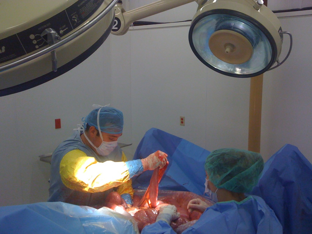
Cultivated Autologous Limbal Epithelial Cell (CALEC) surgery represents a groundbreaking advancement in the field of ocular health, specifically for patients suffering from corneal damage. This innovative procedure involves a meticulous process of harvesting stem cells from a healthy eye, which are then cultivated to create a graft that is transplanted into the damaged eye. By restoring the corneal surface, CALEC surgery offers renewed hope for individuals who have been previously deemed untreatable due to severe limbal stem cell deficiency, often resulting from traumatic injuries or chemical burns.
The procedure showcases the significant potential of stem cell therapy in eye treatments, as evidenced by its impressive success rates in clinical trials. With over 90 percent effectiveness in restoring corneal surfaces in treated patients, CALEC surgery exemplifies the convergence of cutting-edge research and real-world application. The rigorous quality standards for the grafts developed from limbal epithelial cells indicate progress in eye transplant innovations, promising a new avenue for those affected by debilitating corneal conditions.
Clinical Trials and the Future of Limbal Epithelial Cells
The recent clinical trial investigating CALEC therapy highlights the importance of scientific research in bringing innovative eye treatments to fruition. Conducted at Mass Eye and Ear, the trial involved a diverse group of patients whose corneal injuries prohibited traditional treatments like corneal transplants. By using cultivated limbal epithelial cells from their own bodies, the trial heralded a new era in cornea damage treatment, demonstrating that successful restoration is indeed possible even in challenging cases with limited treatment options.
Looking ahead, the research surrounding CALEC surgery underlines the necessity for additional clinical trials to validate these initial findings. Plans to expand studies to larger populations and consider allogeneic sources of limbal stem cells from cadaveric donors could greatly enhance accessibility for patients with bilateral corneal damage. As the field embraces these advancements, the hope is to achieve regulatory approval that consolidates CALEC’s potential as a standard treatment option across the healthcare continuum.
Future research initiatives will also focus on monitoring longer-term outcomes of visual acuity improvements in participants to build a comprehensive understanding of the effectiveness of CALEC therapy. This ongoing evaluation is vital for establishing solid clinical guidelines that facilitate broader adoption of stem cell treatments in ophthalmology.
The Role of Limbal Stem Cells in Corneal Health
Limbal epithelial cells play a crucial role in maintaining the structural integrity and clarity of the cornea. At the outer edge of the cornea, these cells are responsible for healing and regenerating the corneal surface, particularly in the event of injury or disease. Damage to the limbus can lead to significant ocular discomfort, visual impairment, and even blindness if not addressed. Therefore, finding effective methods to replenish these essential cells, such as through CALEC surgery, is paramount for patients with cornea damage.
The integration of biochemical research with practical clinical applications underscores the significance of limbal stem cells in ocular therapies. Treatments derived from insights gained through rigorous scientific studies have the potential to alleviate the burden experienced by patients suffering from corneal dystrophies. As the understanding of their biology deepens, the strategies to harness these cells for therapeutic purposes evolve, promising improved outcomes in eye care.
Innovations in Eye Transplantation Techniques
With the advent of CALEC surgery, eye transplantation techniques are evolving rapidly, driven by advancements in stem cell therapy and regenerative medicine. Traditional corneal transplants have long been the standard treatment for severe corneal damage, but these procedures often come with limitations, including donor availability and the risks of rejection. The innovative CALEC approach circumvents some of these challenges by utilizing a patient’s own cells, minimizing the chances of adverse reactions and enhancing the likelihood of transplant success.
Moreover, the insights gained from CALEC surgery can influence broader eye transplant innovations. As researchers explore the possibility of expanding treatment to patients with damage in both eyes, the landscape of ocular therapies is poised for transformation. This could usher in a new paradigm where the reliance on donor corneas is diminished, allowing individualized and personalized treatment options that cater to complex corneal injuries, thereby expanding access to restorative therapies.
Visual Acuity Improvements Post-CALEC Surgery
One of the key outcomes of CALEC surgery is its positive impact on visual acuity, which is a critical concern for individuals facing corneal damage. The clinical trial indicated varying improvements in visual acuity amongst patients, with significant recoveries noted at multiple follow-up intervals. The fact that many participants reported enhanced vision quality post-procedure reinforces the notion that stem cell therapy can effectively address even the most drastic corneal impairments.
Continued monitoring of visual outcomes in future CALEC studies will be essential for understanding the full scope of benefits associated with this treatment. As researchers gather more data, they will be better equipped to elucidate the relationship between limbal epithelial cell graft success and overall patient quality of life. This not only serves to highlight the therapeutic potential of CALEC surgery but also emphasizes the need for ongoing support and funding for future eye treatment innovations.
Addressing the Challenges of Cornea Damage
Corneal injuries present a unique challenge within the field of ophthalmology, often requiring urgent intervention to prevent long-term damage. Traditional treatment options, such as corneal transplants, may not be feasible for all patients, especially those who have experienced significant limbal stem cell deficiency. The CALEC method offers a new pathway for treating these challenging cases by focusing on stem cell therapy as a viable solution for restoring the corneal surface.
By addressing the root cause of cornea damage through the regeneration of limbal epithelial cells, CALEC surgery represents a paradigm shift in ophthalmic care. Providing effective treatments tailored to individual patient needs can significantly improve not just visual outcomes, but overall quality of life, illustrating the importance of continual investment in research and innovative clinical methodologies for eye health.
The Safety Profile of CALEC Procedures
An essential aspect of any surgical procedure is its safety profile, and the preliminary results from the CALEC clinical trials appear to show a favorable safety outcome. With no serious adverse events reported among participants, the treatment seems to be a promising option for patients with corneal damage. The only noted adverse event was a minor bacterial infection, which was managed effectively, underscoring the need for proper post-operative care in stem cell-based therapies.
Close monitoring and reporting of any complications during and after CALEC surgery helps build trust in this emerging therapy. As researchers continue to gather data on patient outcomes and experiences, they can refine the techniques used in the procedure to enhance safety and efficacy further. The high safety profile opens avenues for future studies aimed at scaling this treatment approach, highlighting the potential for wider adoption in clinical settings.
Future Directions in Stem Cell Eye Therapies
As the field of stem cell therapy for eye disorders advances, the future is ripe with possibilities for innovative treatments that could transform patient care. The success of CALEC surgery has paved the way for further exploration of stem cell applications in treating other ocular diseases, leading to potential breakthroughs that could significantly improve outcomes for individuals with various types of corneal damage.
Research initiatives focusing on expanding the scope of stem cell therapies will be critical in addressing the diverse needs of patients. By investigating the regeneration capabilities of limbal epithelial cells and their applications in alternative therapies, scientists may uncover solutions to ocular complications that have remained elusive until now. The continued collaboration between research institutions and clinical facilities will be essential in driving these advances forward, ensuring robust therapeutic options are available for those experiencing vision impairments.
The Role of FDA Approval in CALEC Therapy Development
Obtaining FDA approval is a significant milestone for any medical treatment, particularly for innovative therapies like CALEC surgery. The process requires rigorous evaluation of clinical data to demonstrate safety and efficacy, a journey that is essential for translating laboratory successes into real-world applications. With the completion of the initial clinical trial showing promising results, the path toward FDA approval for CALEC therapy becomes a central focus for researchers and clinicians alike.
The potential approval of CALEC would mark a historic achievement in the realm of eye therapies, being one of the first stem cell treatments specifically designed for corneal conditions in the United States. This would not only elevate patient care standards but also encourage further research into regenerative medicine applications across various medical fields. The anticipation surrounding FDA approval reflects the hope that more patients can gain access to groundbreaking therapies that restore vision and improve life quality.
Frequently Asked Questions
What is CALEC surgery and how does it relate to stem cell therapy for the eye?
CALEC surgery, which stands for Cultivated Autologous Limbal Epithelial Cells, involves a revolutionary stem cell therapy that utilizes healthy limbal epithelial cells extracted from a patient’s own eye to repair corneal damage. This innovative technique has demonstrated over 90% effectiveness in restoring the cornea’s surface, providing new hope for patients with previously untreatable cornea damage.
How does CALEC surgery treat cornea damage?
CALEC surgery treats cornea damage by harvesting limbal epithelial cells from a healthy eye through a biopsy. These cells are then expanded into a graft that is transplanted into the damaged eye. This method safely restores the corneal surface, addressing issues caused by limbal stem cell deficiency.
What are the success rates of CALEC surgery in clinical trials?
In clinical trials, CALEC surgery has shown impressive success rates, with complete restoration of the cornea in 50% of patients at the three-month follow-up, increasing to 79% and 77% at 12 and 18 months, respectively. Overall success rates remained high at 93% and 92% when accounting for partial successes.
What are the potential risks associated with CALEC surgery?
While CALEC surgery has a high safety profile and no serious adverse events related to the graft have been reported, minor adverse events such as bacterial infections linked to contact lens use have occurred. Continuous monitoring and future trials will help further clarify the risk factors.
Is CALEC surgery currently available to patients in the United States?
As of now, CALEC surgery is still considered experimental and is not offered at Mass Eye and Ear or any U.S. hospital. Further studies are required to seek federal approval before this innovative treatment can be widely accessed by patients.
How does CALEC surgery compare to traditional cornea transplant procedures?
CALEC surgery offers a novel alternative to traditional cornea transplants, especially for patients with limbal stem cell deficiency. Unlike conventional transplants, which may not be suitable for patients with corneal damage due to limbal cell depletion, CALEC utilizes the patient’s own cells to regenerate the corneal surface, minimizing the risk of rejection.
What future developments can we expect from CALEC surgery research?
Future developments in CALEC surgery research include expanding clinical trials to larger populations, improving manufacturing processes, and potentially developing allogeneic methods using donor limbal stem cells. The overarching goal is to achieve FDA approval, allowing this promising treatment to reach more patients suffering from corneal damage.
| Key Points |
|---|
| Ula Jurkunas performs the first CALEC surgery at Mass Eye and Ear, marking a pivotal moment in eye treatment. |
| The clinical trial involved 14 patients and showed a success rate of over 90% in restoring corneal surfaces after treatment with CALEC. |
| CALEC utilizes stem cells from a healthy eye to regenerate damaged corneal tissue. |
| The procedure demonstrated a high safety profile with no serious adverse events among participants. |
| Future studies aim to expand patient access and explore methods for treating patients with bilateral corneal damage. |
| The research is funded by the National Eye Institute and marks the first human study of stem cell therapy for eye treatment in the U.S. |
Summary
CALEC surgery offers a groundbreaking solution in restoring vision for patients with previously untreatable corneal damage. By utilizing stem cells derived from a healthy eye, this innovative procedure has shown remarkable success rates and highlights the potential for further advancements in eye care. The clinical trials at Mass Eye and Ear underscore the effectiveness and safety of CALEC, paving the way for future studies that may provide broader treatment options for those suffering from corneal injuries.


